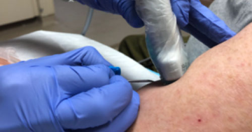
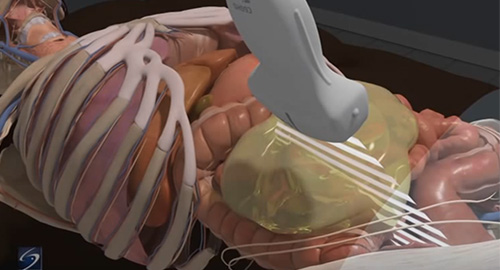
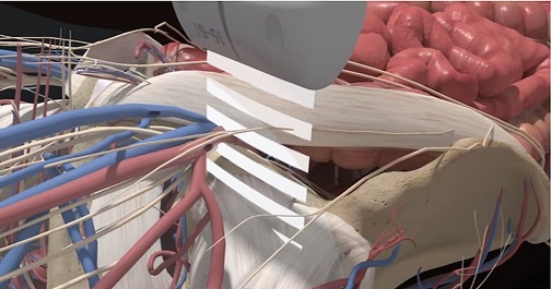
Courses

The following point-of-care ultrasound courses are recommended for you based on what we know about your interests. You can follow the suggested curriculum, or explore other materials at your own pace. To view other available courses, click the courses button at the top of this page.

Femoral Nerve Block

Femoral Nerve Block

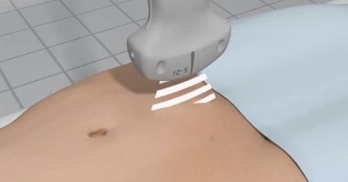
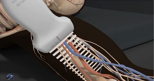
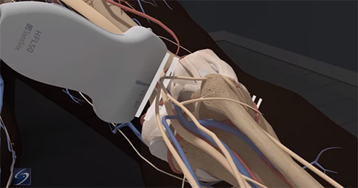
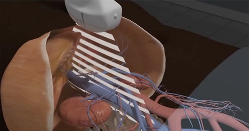
Learn how to avoid common pitfalls by perfecting identification of surrounding anatomical structures, such as the femoral vessels and lymphatic tissues. Understand correct transducer positioning for optimal needle visualization and how to determine sufficient anesthetic spread.

Gallbladder

Gallbladder

The gallbladder is not a fixed organ. Learn the best anatomical landmarks to help you locate and correctly identify the gallbladder and assess for cholecystitis and/or cholelithiasis. Differentiate between hepatic and portal veins and other vessels like ducts and arteries, and learn to spot the symptoms of gallbladder pathology, such as pericholecystic fluid, enlarged bile duct and gallbladder walls, and gallstones.

Interscalene Nerve Block

Interscalene Nerve Block

The Interscalene Nerve Block course is designed for medical professionals utilizing point-of-care ultrasound who want to improve accuracy of nerve blocks using ultrasound guidance. This course covers patient positioning, transducer angling, identification of important vasculature, the location and appearance of the brachial plexus nerves, and the effective position of the needle for analgesic injection.

Ocular

Ocular Ultrasound

The eye can easily be visualized using ultrasound. Pathologies such as retinal detachment, vitreous detachment/hemorrhage, abnormal intracranial pressure, papilledema, and ruptured globe can be identified even by the novice users of ultrasound. Learn which transducers, exam types, and gain/depth settings are ideal for ophthomalogical imaging.

Femoral Nerve Block

Opioid Reduction in Emergency Medicine

This course highlights the basics on various nerve blocks that can be applied to the emergency department setting to reduce the need for opioid prescriptions while treating patients' acute, traumatic pain. Learn more about the growing opioid crisis and steps that can be taken in the emergency department to reduce this epidemic.

Pelvic First Trimester Pregnancy

Pelvic: First Trimester Pregnancy Evaluation

Understand the basic ultrasound views and techniques associated with performing an ultrasound pelvic examination for first trimester pregnancy, surrounding anatomical structures, and proper equipment settings. In addition, support literature, case studies, pathology images, and videos may be included for review. 

Popliteal Sciatic Nerve Block

Popliteal-Sciatic Nerve Block

Review and learn to identify anatomical structures visualized during a popliteal sciatic nerve block with ultrasound guidance; determine the locations of the popliteal artery and vein, and how to locate the perineal and tibial nerve branch, which is ideal for the nerve block placement. 

RUSH

RUSH

The RUSH course is designed for medical professionals with comprehensive knowledge of the core underlying concepts of the RUSH protocol, which includes cardiac, Inferior Vena Cava (IVC), eFAST, lung, aorta, and Deep Vein Thrombosis (DVT) ultrasound examinations. Learn the RUSH protocol for assessing patients in shock. 

Dialysis

Ultrasound-Guided Dialysis (English Only)

Point-of-care ultrasound is a powerful tool for cannulation. As the demographic of the average hemodialysis patient changes, the prevalence of difficult to cannulate vascular accesses increases. This course describes the technique of dialysis cannulation from equipment set-up, patient positioning, and securing the cannula to anatomical variables and pathology.

Ultrasound Guided Paracentesis

Ultrasound-Guided Paracentesis

After completing the Ultrasound-Guided Paracentesis course, viewers should be able to explain the basic outline of ultrasound-guided paracentesis, including: methods for image optimization, ideal equipment selections and settings, identification of abdominal anatomy frequently encountered during paracentesis, and the most common procedural steps involved in performing an ultrasound-guided paracentesis.