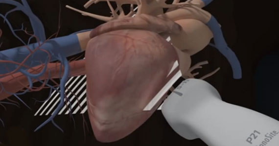
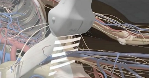
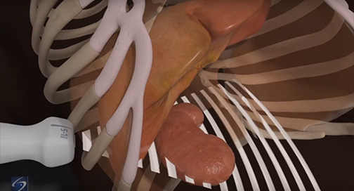
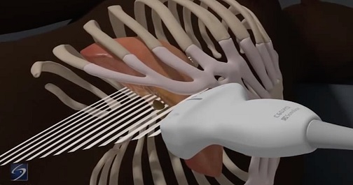
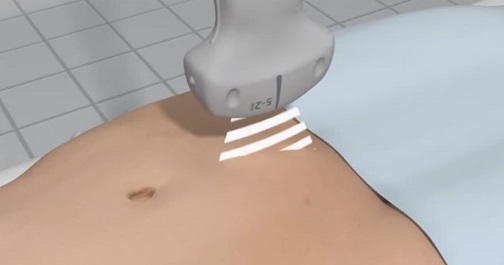
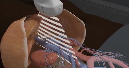
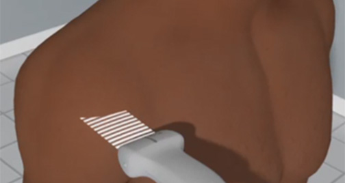
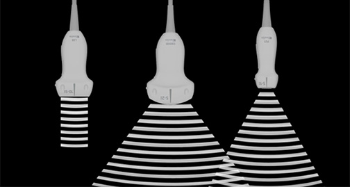
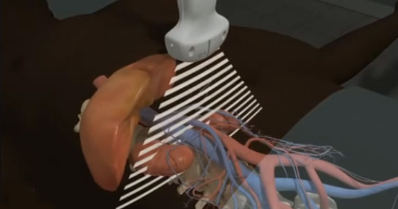
Courses

The following point-of-care ultrasound courses are recommended for you based on what we know about your interests. You can follow the suggested curriculum, or explore other materials at your own pace. To view other available courses, click the courses button at the top of this page.

aorta

Aorta

After completing the aorta ultrasound course, participants should be able to: identify the anatomical structures visualized during the aorta examination, recognize the various types of abdominal aortic aneurysms and perform their associated measurements, and determine the preferred transducer to perform the aorta exam.

Cardiac Imaging 1 Hero

Cardiac Imaging 1

How do you angle the transducer to get a Parasternal Long Axis (PLAX) view of the heart in the Emergency Department? Which kind of transducer should you use? How is the cardiology orientation different? And how, exactly, do you measure fluid responsiveness using the Inferior Vena Cava (IVC)?

Cardiac Imaging 1 introduces these questions (and answers!), and much more. Develop an understanding of how emergency medicine and cardiology applications differ, and test your knowledge of different views of the heart.

Deep Vein Thrombosis

Deep Vein Thrombosis (DVT)

Point-of-care ultrasound is a powerful tool in evaluating a patient for Deep Vein Thrombosis (DVT). This course describes the simplified protocol of assessing the common femoral and popliteal veins for DVT, a method that is well-accepted when used in conjunction with clinical assessments for ruling in/out DVT.

eFAST

eFAST

This valuable exam assesses the trauma patient for internal free fluid collection in the thorax and abdomen. Course participants will learn to identify the internal anatomy seen during the eFAST examination on ultrasound. In addition, students must be able to recognize abnormalities commonly encountered during an eFAST exam, and determine the appropriate transducer for different patient body types.

Gallbladder

Gallbladder

The gallbladder is not a fixed organ. Learn the best anatomical landmarks to help you locate and correctly identify the gallbladder and assess for cholecystitis and/or cholelithiasis. Differentiate between hepatic and portal veins and other vessels like ducts and arteries, and learn to spot the symptoms of gallbladder pathology, such as pericholecystic fluid, enlarged bile duct and gallbladder walls, and gallstones.

Hip

Hip

This course is designed for medical professionals utilizing point-of-care ultrasound who want to build on their hip injection skills. Learn best practices for proximal hamstring, pubic symphysis, and sacroiliac (out of plane technique) injections. Develop an understanding of which surrounding sensitive anatomy to avoid.

Ocular

Ocular Ultrasound

The eye can easily be visualized using ultrasound. Pathologies such as retinal detachment, vitreous detachment/hemorrhage, abnormal intracranial pressure, papilledema, and ruptured globe can be identified even by the novice users of ultrasound. Learn which transducers, exam types, and gain/depth settings are ideal for ophthomalogical imaging.

Pelvic First Trimester Pregnancy

Pelvic: First Trimester Pregnancy Evaluation

Understand the basic ultrasound views and techniques associated with performing an ultrasound pelvic examination for first trimester pregnancy, surrounding anatomical structures, and proper equipment settings. In addition, support literature, case studies, pathology images, and videos may be included for review. 

RUSH

RUSH

The RUSH course is designed for medical professionals with comprehensive knowledge of the core underlying concepts of the RUSH protocol, which includes cardiac, Inferior Vena Cava (IVC), eFAST, lung, aorta, and Deep Vein Thrombosis (DVT) ultrasound examinations. Learn the RUSH protocol for assessing patients in shock. 

Shoulder

Shoulder

When trying to diagnose a partial rotator cuff tear on ultrasound, how should the patient be positioned? Why should you avoid anisotropic artifact during a shoulder scan? What kind of transducer should be used for an obese patient when performing a lateral shoulder injection?

All of these questions, and more, are addressed in the shoulder course. Learn to differentiate normal and abnormal shoulder onscreen anatomy and become familiar with the transducer positioning and angling methods that will help you develop a more accurate ultrasound-guided shoulder injection technique.