Perioperative Ultrasound
This course combines concepts and applications that are encountered in perioperative ultrasound. Take your time and explore sections on airway management, evaluating for gastric content, cardiac views, advanced cardiac imaging, the eFAST exam, the RUSH exam, lung ultrasound and assessment, and ocular pathologies.
As you progress, earn certificates of completion in eFAST, Cardiac Imaging 1, Lung, Ocular, and RUSH with an 80% on higher on each post-test! These passed tests are required prior to taking the Perioperative Ultrasound post-test.
Perioperative Ultrasound: Introduction
Introductory material and disclaimer
Perioperative Ultrasound: Requirements
In order to qualify for the final post-test on Perioperative Ultrasound, you must get an 80% or higher on all post-tests within the sections below.
Airway Management
View these videos about airway management, which can be applied to the perioperative setting.
Lung Ultrasound
Lung ultrasound is a new and exciting frontier for point-of-care imaging. Recently, studies have shown that air, once considered a hindering artifact on ultrasound scans, can be used to identify specific lung pathologies, like pneumonia. This course reviews lung ultrasound as it relates to the point-of-care market.
RUSH Exam
The RUSH course is designed for medical professionals with comprehensive knowledge of the core underlying concepts of the RUSH protocol, which includes cardiac, Inferior Vena Cava (IVC), eFAST, lung, aorta, and Deep Vein Thrombosis (DVT) ultrasound examinations. Learn the RUSH protocol for assessing patients in shock.
Cardiac Imaging 1
How do you angle the transducer to get a parasternal long axis view of the heart in the Emergency Department? Which kind of transducer should you use? How is the cardiology orientation different? And how, exactly, do you measure fluid responsiveness using the Inferior Vena Cava (IVC)? Cardiac 1 introduces these questions (and answers!), and much more. Develop an understanding of how emergency medicine and cardiology applications differ, and test your knowledge of different views of the heart.
Cardiac Imaging 2
View these videos of advanced cardiac imaging. Topics covered include stroke volume determination, transesophageal echocardiography, Doppler principles, bedside valvular assessment, and advanced right ventricular assessment.
Ocular Ultrasound
The eye can easily be visualized using ultrasound. Pathologies such as retinal detachment, vitreous detachment/hemorrhage, abnormal intracranial pressure, papilledema, and ruptured globe can be identified even by the novice users of ultrasound. Learn which transducers, exam types, and gain/depth settings are ideal for ophthomalogical imaging.
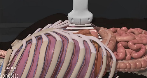
eFAST Exam
This valuable exam assesses the trauma patient for internal free fluid collection in the thorax and abdomen. Course participants will learn to identify the internal anatomy seen during the eFAST examination on ultrasound. In addition, students must be able to recognize abnormalities commonly encountered during an eFAST exam, and determine the appropriate transducer for different patient body types.
Gastric Content
View these videos on evaluating gastric content, which can be applied to the perioperative setting.